
RAISING AWARENESS THROUGH EDUCATION
PROMOTING PREVENTION & EARLY DETECTION CAMPAIGNING FOR CHANGE
SKCIN: THE KAREN CLIFFORD SKIN CANCER CHARITY / REGISTERED CHARITY: 1150048
WE NEED YOUR HELP! PLEASE HELP US TO STOP SKIN CANCER TAKING MORE LIVES. WE ARE HUGELY GRATEFUL FOR YOUR SUPPORT.
Non-Melanoma Skin Cancers
about each type of skin cancer and what signs of change you should look out for - EARLY DETECTION IS VERY IMPORTANT!


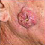
Basal Cell Carcinoma
Basal Cell Carcinoma (BCC) is the most common type of skin cancer. It grows slowly over months and years and may damage nearby tissues and organs if left untreated.
- Basal Cell Carcinoma (BCC) is the most common type of skin cancer.
- BCC represents approximately 80 percent of all diagnosed skin cancers.
- BCC's are uncontrolled, abnormal lesions or growths arising from the basal layer of the
skin's epidermis (the outermost layer). - BCC's often look like open sores, pearly/shiny bumps or scars, pink growths, red patches
or dry patches of dry skin. - The majority of BCC's occur on sun exposed areas such as the face, neck and ears.
- BCC's are locally invasive and grow slowly over months and years.
- BCC's rarely spread (metastasize) beyond the original site to anywhere else on the body.
- If not treated properly or neglected BCC's can cause extensive local invasion and disfigurement.
- Only in extremely rare cases can BCC spread and become life threatening.
- The majority of BCC's are caused by over-exposure to UV from the sun and/or sunbeds.
- Between 70,000 to 125,000 new cases are diagnosed each year in the UK.
- An estimated 2.8 million cases of BCC are diagnosed each year in the US.

There are five warning signs that indicate the possibility of Basal Cell Carcinoma.
However, often two or more of the five warning signs can be present in a single site.
Some of these warning signs can resemble non-cancerous skin conditions such as eczema or psoriasis, it is therefore important to have any abnormalities checked by a skin specialist.
All images used are to help you identify any abnormalities, but all skin cancers can vary in size shape and colour, and many can be very small, so it is important to acknowledge that these should be used as a guideline only. If you notice any of the following warning signs, feel worried or unsure about any change in your skin, consult your doctor immediately.

1: An open, non-healing sore
This is a common warning sign of an early Basal Cell Carcinoma
A persistent sore that oozes, bleeds, crusts for weeks
The sore sometimes heals, but re-opens

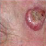
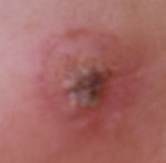
2: A pinkish growth
Growths often have an elevated, rounded border
They may or may not have a crusted indentation to the centre
As the growth enlarges, tiny blood vessels can appear on the surface

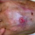
3: Red patch or irritated area of skin
Sometimes patches can crust and itch
They can persist with no evident discomfort
They frequently occur on the face, shoulders, chest, legs and arms

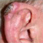
4: A shiny Nodule or bump
These are often pearly or translucent
They can also appear pink, red or white
On darker skinned individuals they can be tan, brown or black

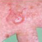
5: A scar-like patch
The area looks like a scar with undefined borders
The scar-like area often looks white, yellow and is waxy
This could indicate an invasive BCC that is much larger than it appears from the surface
What causes Basal Cell Carcinoma?
The major cause of Basal Cell Carcinoma is exposure to UV radiation from the sun and/or sunbeds, which is why the majority of all BCC cases are found on frequently exposed areas of the body such as the face, neck, ears, chest, shoulders, back and scalp.
On rare occasions BCC's can be found on unexposed areas and in a fewer cases, open sores, chronic skin conditions, burns, scars, tattoos and vaccinations, in addition to contact with arsenic and exposure to radiation.
Who is at risk of developing Basal Cell Carcinoma?
Anyone with a history of sun exposure can get Basal Cell Carcinoma, although those with higher levels of sun exposure and/or fair skinned individuals are generally at greater risk.
Older people are usually affected more by this skin cancer, but the age is steadily decreasing with a higher reported number of those being diagnosed in their 20's and 30's.
An increasing number of women are also being diagnosed with BCC, which has previously been more prevalent in men - possibly due to outdoor occupations and leisure pursuits.
Following a specialist examination, the diagnosis of BCC is confirmed with a biopsy.
To perform the biopsy the skin is numbed with local anaesthesia. A piece of tissue is then removed and examined under a microscope to confirm the diagnosis. If tumour cells are present, treatment is required.
There are a number of highly effective treatments for Basal Cell Carcinoma, which treatment is right for a specific patient depends on the type, size, location, and depth of penetration of the tumour as well as the patient's age and general health. Treatment can almost always be performed on an outpatient basis.
5-year cure rate for primary BCC
Mohs Micrographic Surgery 99%
Excisional surgery 89.9%
Cryotherapy 92.5%
Curettage and cautery 92.3%
Radiotherapy 91.3%
Reference: Rowe DE, Carroll RJ, Day CL Jr. Long-term recurrence rates in previously untreated (primary) basal cell carcinoma: implications for patient follow-up.
Excisional Surgery:
A local anaesthetic surgery with predetermined margins - also known as Wide Local Excision (WLE). This is highly effective treatment for BCC. For well defined BCC < 2cm, excision margins of 4mm will result in a 95% clearance rate.
A scalpel is used to remove the entire growth and surrounding border of apparently normal or healthy skin. The wound is then closed with stitches. The procedure takes 30-60 minutes depending on the complexity of the cancer but is usually straightforward.
The excised tissue is sent for microscopic examination to confirm that all cancerous cells have been removed.
Stitches are usually in place for 5-7 days.
Recurrent BCC require wider excisional surgery or Mohs micrographic surgery.
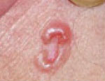
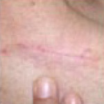
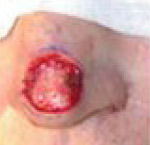
Example of Wide Local Excision
of Bases Cell Carcinoma. Patient: John Holmes
Mohs Micrographic surgery
Mohs micrographic surgery is most commonly used to remove basal cell carcinomas, but can sometimes be used to remove other tumours. Using a scalpel the visible tumour is removed along with a very thin layer of tissue surrounding it. This layer is then checked immediately and thoroughly under a microscope. If the tumour is still present in or around the surrounding tissue, the procedure is repeated until the area is tumour-free.
Mohs Micrographic Surgery saves the greatest amount of healthy tissue, and has the highest overall cure rate of any treatment for basal cell carcinoma and squamous cell carcinoma.
It is frequently used on tumours that have recurred, are located in hard-to-treat, or critical areas such as around the eyes, nose, lips, and ears, as well as the neck, hands and feet.
After surgery, the wound may be allowed to heal naturally or if necessary be reconstructed using plastic surgery methods.
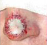
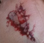
Example of Mohs Micrographic Surgery:
Removal of Basal Cell Carcinoma. Patient: Richard Clifford


Curettage and Cautery
Under local anaesthetic, the cancerous growth is scraped off with a curette (a sharp, ring-shaped instrument). An electrocautery needle producing heat then destroys residual tumour and controls
bleeding. This treatment may be repeated a few times to ensure that all cancer cells are eliminated. It can produce cure rates similar to those of surgical excision, but is not as useful for aggressive skin
cancers or those located in high-risk or difficult sites.
Cryotherapy
Cryotherapy means 'treatment using low temperature', and refers to the removal of skin lesions by freezing them. Many superficial benign lesions can be treated with cryotherapy, but it is most commonly used to remove actinic keratoses, seborrhoeic keratoses and other small skin cancers such as basal cell carcinomas, superficial squamous cell carcinoma and Bowen's disease.
The tumour tissue is destroyed by freezing it with liquid nitrogen, this is done by using a spray device or cotton-tipped applicator. Cryotherapy does not normally require a local anaesthetic and it involves no cutting or bleeding, the procedure itself lasting a matter of seconds.
The area of frozen skin will turn white, thawing to normal skin temperature within a couple of minutes. The procedure may be repeated to ensure the eradication of all malignant cells. Swelling, redness and blistering can occur following treatment. The growth then scabs and falls off within weeks. The treated area will usually look normal once healed, but some scarring can or discoloration can occur particularly on the legs or on darker skinned patients. (It is important not to pick the wound during healing). This treatment has a lower overall cure rate than surgical methods but may be a treatment of choice for some patients.
Radiotherapy
During Radiotherapy X-ray beams are directed at the tumour, with no need for cutting and no local anaesthetic. To ensure destruction of the tumour a series of treatments are often required and administered several times a week for up to four weeks, or sometimes daily for one month. Cure rates range from around 85 to 95 percent, but the technique can involve long-term cosmetic problems and radiation risks, as well as multiple visits. For these reasons, this Radiotherapy is generally used for patients for whom surgery is not advised, such as the elderly or those in poor health and for tumours that are particularly hard to treat surgically.
Photodynamic Therapy (PDT)
Photodynamic therapy or PDT is a relatively new type of treatment and an alternative to surgery in some cases. It is basically treatment with a chemical that makes the skin cells sensitive to light and
when the area to be treated is exposed to laser light, the cells die off.
It is best used on thinner skin cancers, in cases where you would need a lot of surgery and can be especially useful for growths on the face and scalp. It is not suitable for deeper skin cancers because the light cannot penetrate far enough into the skin.
PDT is not recommended for squamous cell skin cancers because there is too high a risk of the cancer coming back. PDT is now available on the NHS for Bowen�s disease, basal cell skin cancers and actinic keratosis (solar keratosis).
To have this treatment, you first have the crust and scale removed from your skin tumour, you then have a cream that contains light sensitising chemical applied to the skin cancer and the surrounding area. Sometimes, you may have this chemical as a tablet or injection. After the drug has been absorbed, you will have a strong light shone on to the treated area for up to 45 minutes. The light will kill any cell that has absorbed the drug. This treatment is likely to be repeated more than once.
Redness and swelling are common side effects. After treatment, patients become locally photosensitive for 48 hours where the light sensitising chemical was applied, and must avoid the sun.
Topical Medications
Imiquimod is FDA-approved only for superficial BCCs, with cure rates generally between 80 and 90 percent. (Aldara) is the cream that is licensed for the treatment of superficial BCC. This cream is useful for low-risk BCC in non-critical sites. Applied 5 times a week for 6 weeks, it evokes an inflammatory reaction. Assessment of response takes place after a further 12 weeks.
For selected BCC, this is a good option with good cure rates but long term follow up data is sparse. It is the first in a new class of drugs that work by stimulating the immune system.
5-Fluorouracil (5-FU) is also FDA-approved for superficial BCCs, with similar cure rates to imiquimod. The 5% liquid or ointment is gently rubbed into the tumour twice a day for three to six weeks.
Trials with more invasive BCCs are under way for both imiquimod and 5-FU. Side effects are variable, and some patients do not experience any discomfort, but soreness can occur and redness, irritation and inflammation are predictable.
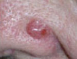
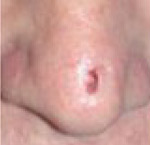
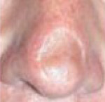
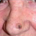
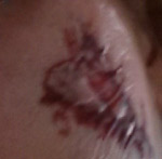
Example of BCC Operation on the Nose
Removal of Basal Cell Carcinoma. Patient: John Holmes
Prevention is better than cure.
Whilst most skin cancers and precancers are curable if detected early, prevention is always better than cure. 80-90% of all skin cancers can be avoided by ensuring that appropriate precautions are
taken during time spent in the sun.
Always follow the five S's of sun safety below and make sure you stick to these golden rules:
- Avoid tanning and never allow your skin to burn
- Never use indoor tanning devices
- Examin your skin thoroughly at least once a month
- If you are worried or uncertain about any abnormality, seek immediate professional advice
For those that have suffered from a precancer, or skin cancer, particular care and attention to covering up and shading from the sun is very important as well as thoroughly checking your skin more frequently for signs of recurrence or developments of new abnormalities.
The Five S's of Sun Safety:

SLIP on a T-shirt
Always keep shoulders covered
as they can easily burn in the sun

SLOP on good quality sunscreen
Always use a sunscreen with a minimum SPF of 30 with broad spectrum UVA protection
Apply generously 20 mins before going outside and reapply at least every 2 hours

SLAP on a broad brimmed hat
Wear a legionnaire or broad-brimmed hat to help shade the face, neck and ears
which can easily burn in the sun.

SLIDE on quality sunglasses
Wear quality (preferrably wrap-around) sunglasses to help protect
your eyes that can be damaged by the sun.

SHADE - Seek shade whenever possible
Always seek shade, particularly between 11am and 3pm when the sun's
UV penetration is strongest.


Squamous Cell Carcinoma
Squamous Cell Carcinoma (SCC) is less common but grows faster. It may spread to other parts of the body and if left untreated can be deadly.
- Squamous Cell Carcinoma (SCC) is the second most common type of non-melanoma skin cancer.
- SCC is most commonly found in older people, but the popularity of sunny holidays and recreational
use of sunbeds means that we are seeing them in younger age groups. - SCC is an uncontrolled growth of abnormal squamous cells in the skin's epidermis (most outer layer) and is a cancer of the keratinocytes of the skin.
- Squamous Cell Carcinoma is mainly caused by cumulative UV exposure.
- SCC is usually found on areas of skin often exposed to the sun, typically the face, ears, lips, mouth, hands, arms and legs but can appear anywhere on the body.
- The appearance varies but is usually a scaly lump, nodule, ulcer or non-healing sore or wart that is elevated with a central depression and may crust and bleed.
- They often start as small hard white or skin-coloured lumps in the skin that grow at a variable rate.
- SCC's have the ability to spread to other parts of the body, but do not often do so.
- SCC can become disfiguring if left untreated.
- In severe cases SCC can spread to local lymph nodes or around the body and result in death.

- SCC is usually found on areas of skin often exposed to the sun, typically the face, ears, lips, mouth, hands, arms and legs but can appear anywhere on the body.
- The appearance varies but is usually a scaly lump, nodule, ulcer or non-healing sore or wart that is elevated with a central depression and may crust and bleed.
- They often start as small hard white or skin-coloured lumps in the skin that grow at a variable rate.
Below are 4 warning signs that indicate the possibility of Squamous cell Carcinoma
Sometimes two or more of these warning signs can be present in a single site. All images used here are to help you identify any abnormalities, but all skin cancers can vary in size shape and colour and texture, so it is important to acknowledge that these should be used as a guideline only. If you notice any of the following warning signs, feel worried or unsure about any change in your skin, consult your doctor immediately.

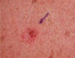
1: A persistent Scaly patch
Often red in colour, with uneven borders
Look out for a scaly patch of skin that won't heal
Often these patches will crust and bleed

2: An elevated growth with central depression
Growths are raised with an indentation to the centre
Growths can sometimes bleed
They can grow rapidly in size

3: Open sore
Look out for an open sore-like wound
A sore that constantly crusts and bleeds
A persistent sore that won't heal over weeks

4: A wart-like growth
Look out for a growth that resembles a wart
A growth that crusts and sometimes bleeds
A wart-like growth that won't heal or respond to treatment
What causes Squamous Cell Carcinoma?
Squamous Cell Carcinoma is mainly caused by chronic and cumulative exposure to UV radiation from the sun and or sunbeds. SCC can also arise in skin injuries such as scars, burns, ulcers, sores.
Occasionally, a squamous cell carcinoma may be caused by exposure to certain substances used in industry, such as tar and can sometimes develop in patients who are on drugs that suppress the immune system.
Who is at risk of developing Squamous Cell Carcinoma?
Anyone with a history of sun exposure can get Squamous Cell Carcinoma, although those with higher levels of sun exposure and/or fair skinned individuals are generally at greater risk.
SCC is often found in older individuals but is on the increase in younger generations due to obsessions with tanning and the use of sunbeds. Research in the Netherlands has also shown that smoking more than triples the risk of developing squamous cell Carcinoma.
Squamous cell carcinomas are more than twice as frequent in men as in women.
Outdoor workers or those who spend extensive leisure time in the sun are also at greater risk.
Anyone who has had a basal cell carcinoma is also more likely to develop an squamous cell carcinoma, as is anyone with an inherited, highly UV-sensitive condition such as xeroderma pigmentosum.
If detected at an early stage and removed promptly Squamous Cell Carcinomas are almost always curable and cause minimal damage and discomfort. In more serious cases however - where a Squamous Cell Carcinoma has been left untreated, the tumour can penetrate the underlying tissue and become disfiguring. It is also possible for Squamous Cell Carcinoma, if left untreated to metastasise and spread to organs and be fatal. It is therefore extremely important to seek professional advice with any suspicious growth.
A biopsy of the suspicious growth will be examined under a microscope and a diagnosis will be made. There are a number of highly effective treatments for Basal Cell Carcinoma, which treatment is right for a specific patient depends on the type, size, location, and depth of penetration of the tumour as well as the patient's age and general health. Treatment can almost always be performed on an outpatient basis.
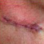
Excisional Surgery
A local anaesthetic surgery with predetermined margins - also known as Wide Local Excision (WLE).
A scalpel is used to remove the entire growth and surrounding border of apparently normal or healthy skin. The wound is then closed with stitches. The procedure takes 30-60 minutes depending on the complexity of the cancer but is usually straightforward.
The excised tissue is sent for microscopic examination to confirm that all cancerous cells have been removed. Stitches are usually in place for 5-7 days.
Recurrent SCC will require wider excisional surgery or Mohs micrographic surgery.
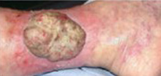
Example of Surgical Excision
of Squamous Cell Carcinoma on the cheek.

Mohs Micrographic surgery
Mohs micrographic surgery is most commonly used to remove basal cell carcinomas, but can sometimes be used to remove other tumours. Under local anaesthetic the visible tumour is removed with a scalpel along with a very thin layer of tissue surrounding it. This layer is then checked immediately and thoroughly under a microscope. If the tumour is still present in or around the surrounding tissue, the procedure is repeated until the area is tumour-free.
Mohs Micrographic Surgery saves the greatest amount of healthy tissue, and has the highest overall cure rate of any treatment for basal cell carcinoma and squamous cell carcinoma.
It is frequently used on tumours that have recurred, are located in hard-to-treat, or critical areas such as around the eyes, nose, lips, and ears, as well as the neck, hands and feet.
After surgery, the wound may be allowed to heal naturally or if necessary be reconstructed using plastic surgery methods.
Example of Mohs Micrographic Surgery:
Removal of Basal Cell Carcinoma. Patient: Richard Clifford
Curettage and Cautery
Under local anaesthetic, the cancerous growth is scraped off with a curette (a sharp, ring-shaped instrument). An electrocautery needle producing heat then destroys residual tumour and controls bleeding. This treatment may be repeated a few times to ensure that all cancer cells are eliminated.
It can produce cure rates similar to those of surgical excision, but is not as useful for aggressive skin cancers or those located in high-risk or difficult sites.
Cryotherapy
Cryotherapy means 'treatment using low temperature', and refers to the removal of skin lesions by freezing them. Many superficial benign lesions can be treated with cryotherapy, but it is most commonly used to remove actinic keratoses, seborrhoeic keratoses and other small skin cancers such as basal cell carcinomas, superficial squamous cell carcinoma and Bowen's disease.
The tumour tissue is destroyed by freezing it with liquid nitrogen, this is done by using a spray device or cotton-tipped applicator. Cryotherapy does not normally require a local anaesthetic and it involves no cutting or bleeding, the procedure itself lasting a matter of seconds.
The area of frozen skin will turn white, thawing to normal skin temperature within a couple of minutes. The procedure may be repeated to ensure the eradication of all malignant cells.
Swelling, redness and blistering can occur following treatment.
The growth then scabs and falls off within weeks.
The treated area will usually look normal once healed, but some scarring can or discoloration can occur particularly on the legs or on darker skinned patients. (It is important not to pick the wound during healing). This treatment has a lower overall cure rate than surgical methods but may be a treatment of choice for some patients.
Radiotherapy
During Radiotherapy X-ray beams are directed at the tumour, with no need for cutting and no local
anaesthetic. To ensure destruction of the tumour a series of treatments are often required and administered several times a week for up to four weeks, or sometimes daily for one month.
Cure rates range from around 85 to 95 percent, but the technique can involve long-term cosmetic problems and radiation risks, as well as multiple visits. For these reasons, this Radiotherapy is generally used for patients for whom surgery is not advised, such as the elderly or those in poor health and for tumours that are particularly hard to treat surgically.
Prevention is better than cure.
Whilst most skin cancers and precancers are curable if detected early, prevention is always better than cure. 80-90% of all skin cancers can be avoided by ensuring that appropriate precautions are
taken during time spent in the sun.
Always follow the five S's of sun safety below and make sure you stick to these golden rules:
- Avoid tanning and never allow your skin to burn
- Never use indoor tanning devices
- Examin your skin thoroughly at least once a month
- If you are worried or uncertain about any abnormality, seek immediate professional advice
For those that have suffered from a precancer, or skin cancer, particular care and attention to covering up and shading from the sun is very important as well as thoroughly checking your skin more frequently for signs of recurrence or developments of new abnormalities.
The Five S's of Sun Safety:
SLIP on a T-shirt
Always keep shoulders covered
as they can easily burn in the sun
SLOP on good quality sunscreen
Always use a sunscreen with a minimum SPF of 30 with broad spectrum UVA protection
Apply generously 20 mins before going outside and reapply at least every 2 hours
SLAP on a broad brimmed hat
Wear a legionnaire or broad-brimmed hat to help shade the face, neck and ears
which can easily burn in the sun.
SLIDE on quality sunglasses
Wear quality (preferrably wrap-around) sunglasses to help protect
your eyes that can be damaged by the sun.
SHADE - Seek shade whenever possible
Always seek shade, particularly between 11am and 3pm when the sun's
UV penetration is strongest.
THE EARLY DETECTION OF SKIN CANCER CAN REDUCE YOUR RISK OF POTENTIAL DISFIGURMENT OR EVEN DEATH.
Make sure you check your skin thoroughly at least once a month for signs of change.
Remember what to look out for when checking your skin - download and print off our early detection fact sheet.
DOWNLOAD NOWREMEMBER: If you are worried or uncertain about any suspicious patch, lump, mole or lesion - go to your GP and seek professional advice from a skin specialist or dematologist immediately.
REMEMBER: PREVENTION IS BETTER THAN CURE!
Always use the
Five S's of Sun
Safety & never allow
your skin to burn!